


Gifting Made Simple
Give the Gift of ChoiceClick below to purchase a Prairie Mall eGift Card that can be used at participating retailers at Prairie Mall.Buy Gift CardHome
Atlas of Sectional Anatomy: Understanding the Anatomical Aspects Thorax, Abdomen and Pelvis
Coles
Loading Inventory...
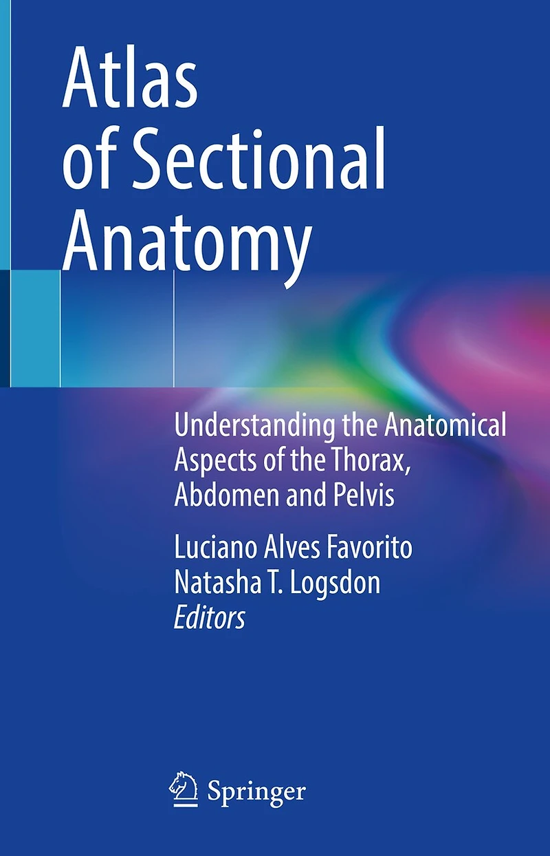
Atlas of Sectional Anatomy: Understanding the Anatomical Aspects Thorax, Abdomen and Pelvis in Grande Prairie, AB
Current price: $262.95
Coles
Atlas of Sectional Anatomy: Understanding the Anatomical Aspects Thorax, Abdomen and Pelvis in Grande Prairie, AB
Current price: $262.95
Loading Inventory...
Size: Hardcover
*Product information and pricing may vary - to confirm current pricing, availability, shipping, and return information please contact Coles. In the event of a pricing discrepancy, the retailer's price will apply.
Sectional anatomy is a valuable resource for understanding and interpreting imaging exams, specially computed tomography (CT) and magnetic resonance imaging (MRI). Thus, health professionals should have a solid anatomical knowledge to properly evaluate such exams during clinical assessments of cardiac, thoracic, abdominal, proctologic, gynecological and urological diseases. The chapters in this book describe the thoracic anatomy, the abdominal wall, retroperitoneal space, and the male and female pelvis. Sectional images of cadaveric material illustrate the thoracic and the abdominal cavities, kidney, ureter, prostate, penis and other male and female organs. The images and descriptions build familiarity with the anatomical traits and can be applied in the fields of urology, gynecology, proctology, radiology and surgery. This work appeals to a wide range of readers, from health professionals to residents and students of different medical specialties.
Sectional anatomy is a valuable resource for understanding and interpreting imaging exams, specially computed tomography (CT) and magnetic resonance imaging (MRI). Thus, health professionals should have a solid anatomical knowledge to properly evaluate such exams during clinical assessments of cardiac, thoracic, abdominal, proctologic, gynecological and urological diseases. The chapters in this book describe the thoracic anatomy, the abdominal wall, retroperitoneal space, and the male and female pelvis. Sectional images of cadaveric material illustrate the thoracic and the abdominal cavities, kidney, ureter, prostate, penis and other male and female organs. The images and descriptions build familiarity with the anatomical traits and can be applied in the fields of urology, gynecology, proctology, radiology and surgery. This work appeals to a wide range of readers, from health professionals to residents and students of different medical specialties.
You May Also Like










Find at Prairie Mall in Grande Prairie, AB
Visit at Prairie Mall in Grande Prairie, AB